
Figure 4.
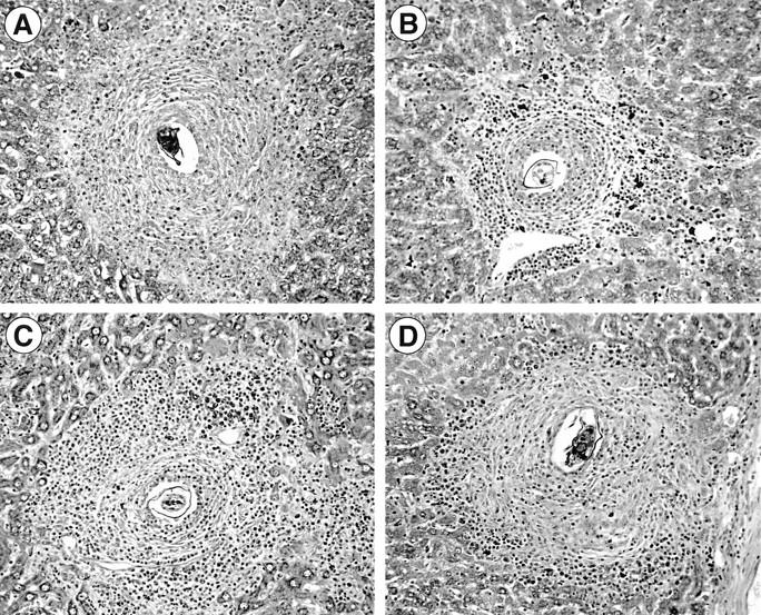
Photomicrographs of representative hepatic granulomas from WT control versus KO animals. The granulomas shown are from 8-wk (A) and 16-wk (B) infected C57BL/6J mice, and from 16-wk infected μMT animals (C) and 16-wk infected FcRγ KO mice (D). Original magnification of Giemsa stain, ×200.