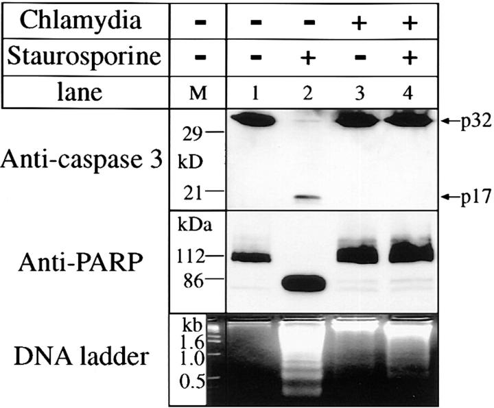
Figure 3.
Effect of chlamydial infection on caspase 3 processing, PARP cleavage, and DNA fragmentation. HeLa cells with (lanes 3 and 4) or without (lanes 1 and 2) chlamydial infection and with (lanes 2 and 4) or without (lanes 1 and 3) staurosporine (1 μM) treatment were lysed for Western blot analysis using antibodies against caspase 3 (top) or PARP (middle). The caspase 3 and PARP antibody staining was developed with a secondary antibody conjugated to horseradish peroxidase followed by visualization using an ECL as described in Materials and Methods. A 3% agarose gel was used for the DNA ladder assay and ethidium bromide was used to visualize the DNA bands (bottom).

