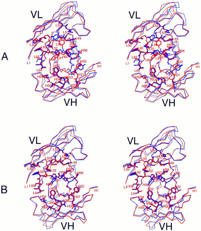
Figure 1.

Antigen combining site of free and complexed HuLys and D1.3, wall-eyed stereo views. (A), unliganded forms. Molecule 2 of free HuLys (14) is shown superposed on free D1.3 Fv. (B), Complexed forms. Molecule 1 of the HuLys–lysozyme complex crystal form is shown superposed on D1.3 Fv from the D1.3–hen lysozyme complex. Lysozyme has been removed from the illustration. Superpositions were performed with QUANTA, using the Cα atoms of antigen-contacting residues. A list of lysozyme-contacting residues in D1.3, which match those seen in the HuLys– lysozyme complex, is given in reference 35. The structures are shown from analogous “antigen eye views” looking directly at the antigen combining site of each antibody. For simplicity, residues that do not directly contact antigen are drawn as a Cα trace, whereas antigen-contacting residues are fully drawn. HuLys, red; D1.3, purple. The illustration was drawn using the program MOLSCRIPT (46).
