
Figure 8.
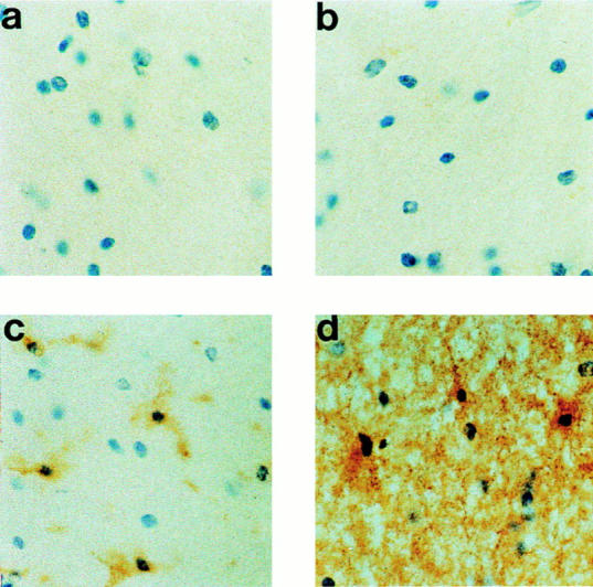
Immunodetection of perforin in normal human brain tissue. Frozen tissue sections were stained (a) using an irrelevant antibody (OX23, mouse antifactor H) or with mouse antibody against (b) perforin, (c) HLA class II (clone LN3), or (d) CD44 (clone BRIC222) DAB/immunoperoxidase protocol and were counterstained using hematoxylin. No specific staining for perforin was obtained in normal surgical brain, whereas quiescent microglia and astrocytes were identified respectively by the HLA class II and CD44 stainings (original magnification: ×500).