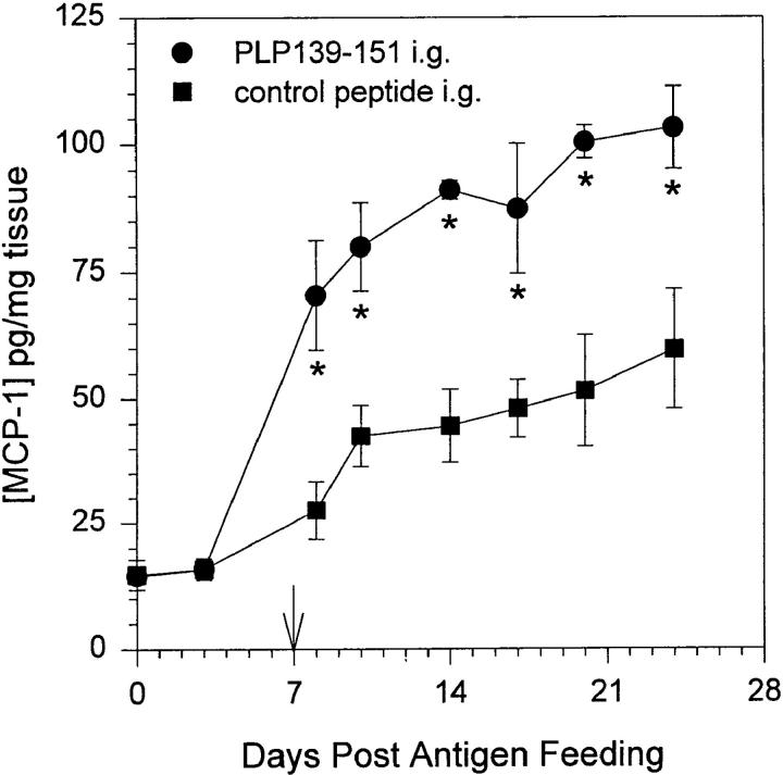
Figure 1.
MCP-1 production in intestinal mucosa after antigen feeding. Mice were fed either PLP139–151 or control peptide and 7 d later were primed for EAE induction by injection of PLP139–151 in CFA (arrow). Intestinal mucosa was harvested from representative mice at various timepoints after antigen feeding, homogenized, and assayed for the presence of MCP-1 protein by ELISA. The data represent the mean MCP-1 levels from duplicate assay samples. The data are representative of three identical experiments. Asterisk, P <0.05.

