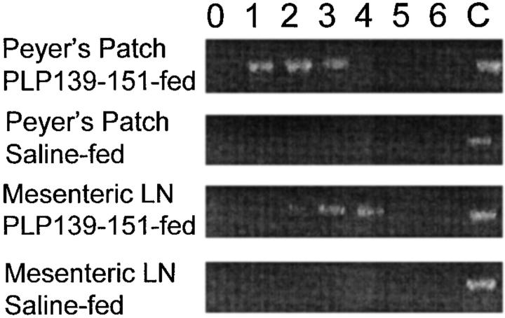
Figure 2.
Detection of MCP-1 mRNA expression in mucosal lymphoid tissue after antigen feeding. Mice were fed either PLP139–151 or control peptide and both Peyer's patch and MLNs were harvested and assayed for MCP-1 mRNA expression using RT-PCR. Tissue was harvested at the indicated time points (in days) after oral antigen administration. Control MCP-1 mRNA expression (C) was determined from LPS-stimulated macrophages and used as an internal control for the RT-PCR conditions. Amplified cDNA was electrophoresed through 2% agarose gels and visualized using ethidium bromide. The data are representative of three identical experiments.

