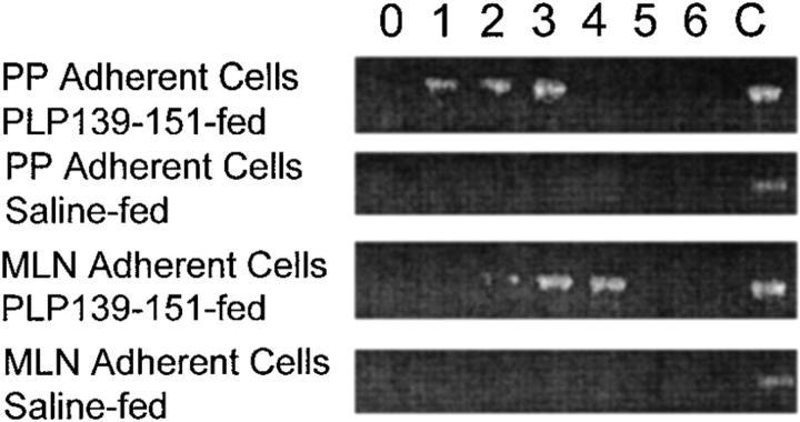
Figure 3.
Detection of MCP-1 mRNA expression in the adherent cell populations of mucosal lymphoid tissue after antigen feeding. Mice were fed either PLP139–151 or control peptide and both Peyer's patch and MLNs were harvested. The adherent cells were isolated by plastic adherence (see Materials and Methods) and assayed for MCP-1 mRNA expression using RT-PCR. Adherent cells were harvested at the indicated time points (in days) after oral antigen administration. Control MCP-1 mRNA expression (C) was determined from LPS-stimulated macrophages and used as an internal control for the RT-PCR conditions. Amplified cDNA was electrophoresed through 2% agarose gels and visualized using ethidium bromide. The data are representative of three identical experiments.

