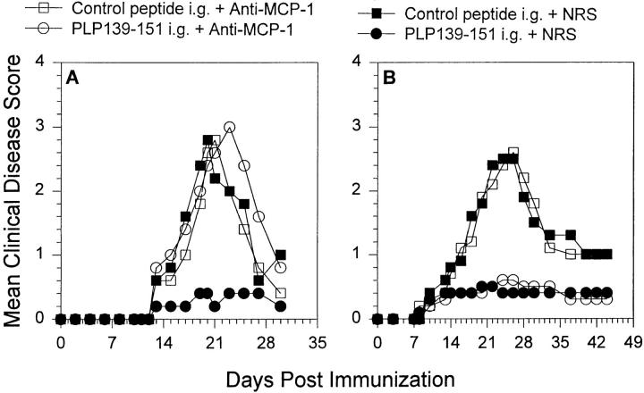
Figure 7.
Il-4 and IL-12 expression in mucosal tissue after oral administration of antigen and in vivo anti–MCP-1 treatment. Mice were fed either PLP139–151 to induce immunologic tolerance or control peptide 7 d before disease induction with PLP139–151 in CFA. Each of those two groups was treated with either 0.5 ml anti–MCP-1 or NRS 2 d before antigen feeding, the day of antigen feeding, and 2 d after antigen feeding (similar to the experiment in Fig. 2). 7 d after disease induction, intestinal mucosa was harvested and analyzed for the presence of IL-4 and IL-12. The data represent either the mean IL-4 or IL-12 levels from duplicate assay samples. The data are representative of two identical experiments.

