
Figure 9.
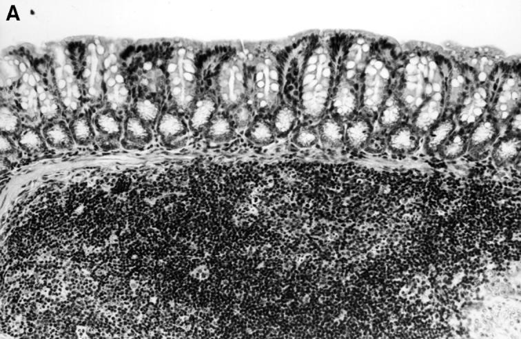
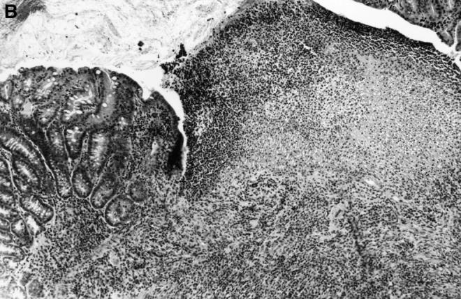
Histopathology of the colon of C3H/HeSnJ scid/scid mice that had been injected with CD4+ T cells from either C3H/HeJBir or C3H/HeJ mice 3 mo before. Before the transfer, CD4+ T cells from both sources were activated for 4 d with cecal bacterial antigen-pulsed APCs. Three months after transfer, the colon of the recipient of the activated C3H/HeJ T cells (left panel) shows normal mucosa overlying a lymphoid follicle, whereas the colon of the recipient of activated C3H/HeJBir T cells (right panel) shows inflammation and a focal ulcer.