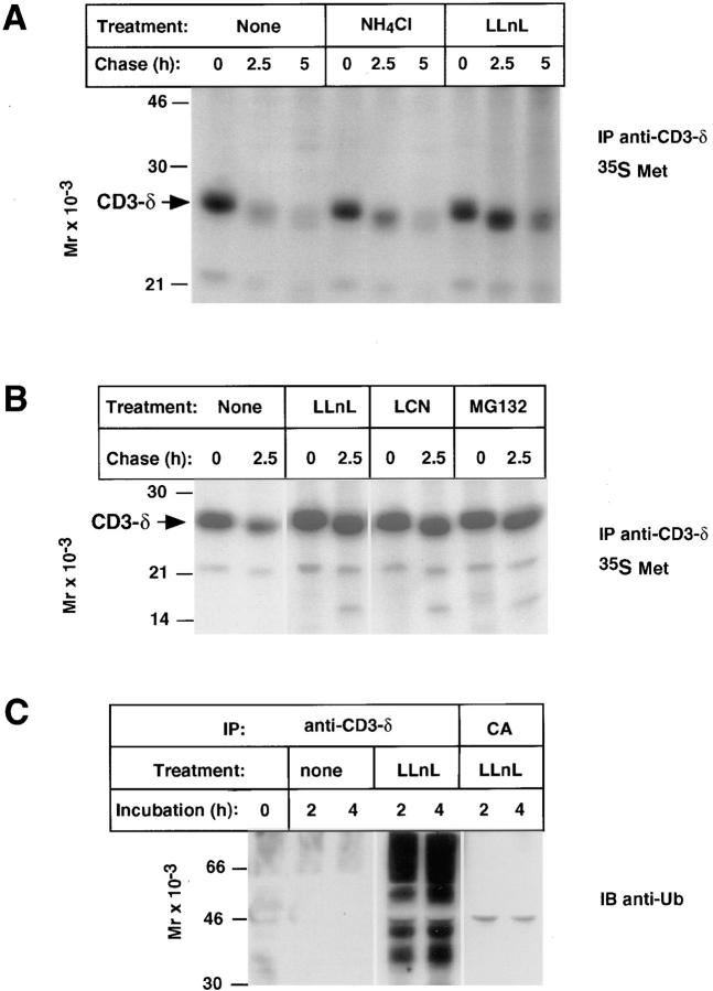
Figure 1.
Proteasomal degradation and ubiquitination of CD3-δ in thymocytes. (A and B) Thymocytes from 12- (A) or 6-wk-old (B) mice were preincubated with the indicated treatments for 1 h at 4°C before labeling for 20 (A) or 30 min (B) at 37°C with [35S]methionine followed by a chase in complete medium with the indicated additions still present. CD3-δ was immunoprecipitated (IP) from cell lysates using an anti–CD3-δ antiserum raised in rabbit (also known as R9; reference 29) and samples were resolved by SDS-PAGE under reducing conditions followed by autoradiography. The position of CD3-δ is indicated. The minor 16-kD band seen at chase points from proteasome-treated samples in B may represent a small amount of deglycosylated CD3-δ. (C) Thymocytes were incubated at 37°C as indicated, followed by immunoprecipitation of cell lysates with either anti–CD3-δ or CA. Immunoblotting (IB) was with polyclonal anti-Ub as previously described (30).

