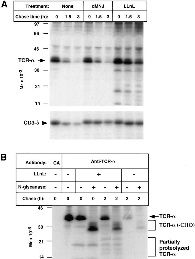
Figure 6.
Degradation of TCR-α in 21.2.2 cells. (A) Cells were labeled and chased in cold medium for the indicated times followed by sequential immunoprecipitation with anti–TCR-α (upper panel) followed by anti–CD3-δ (lower panel). (B) Cells were labeled and chased as indicated and immunoprecipitates were treated with or without N-glycanase. Positions of TCR-α, partially and fully deglycosylated TCR-α, and immunoreactive degradation products are indicated.

