
Figure 1.
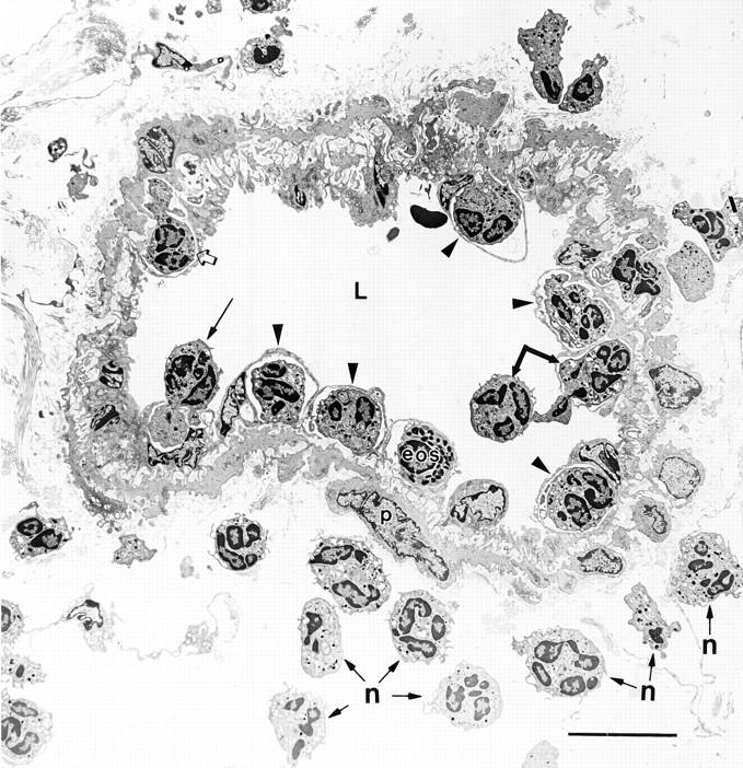
Large venule in guinea pig skin harvested 60 min after intradermal injection of 10−5 FMLP. Many neutrophils and a single eosinophil (eos) are captured at various stages of attachment to and extravasation across vascular endothelium and underlying pericytes (p). Two neutrophils (single joined arrow), one in the lumen and another partway across the endothelium, are tethered together. Another neutrophil (long arrow) has projected a cytoplasmic process into an underlying EC. Other neutrophils (arrowheads) and the eosinophil have crossed the EC barrier, but remain superficial to pericytes, forming dome-like structures that bulge into the vascular lumen. Still another neutrophil (open arrow) that has already crossed the endothelium has extended a process into the basal lamina and indents an underlying pericyte. Other neutrophils (some indicated by n) have crossed both the EC and pericyte barriers and have entered the surrounding connective tissues. L, lumen. Bar, 10 μm.