
Figure 2.
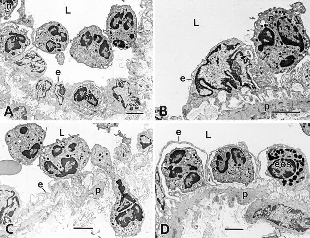
Progression of neutrophil migration across venular endothelium at 60 min after intradermal injection of 10−5 M FMLP. (A) Three neutrophils in the vascular lumen (L) are tethered together at point contacts; two of these also attach to the endothelium (e) at disparate sites. Three additional neutrophils (n) have already crossed the endothelial barrier. (B) An adherent neutrophil (n) projects two separate pseudopods into an EC (e). As confirmed in deeper serial sections, the smaller of these (arrowhead) projects into the EC at a point adjacent to the nucleus, whereas the larger (dominant) pseudopod forms a blunt projection into a thinned region of EC cytoplasm nearby. The EC shows extensive wrinkling of its abluminal surface, except over the nucleus, which is rounded with pinched folds and bulges into the lumen. (C) A chain of three interconnected neutrophils. One of these (elongate cell) has traversed both endothelium (e), basal lamina, and pericyte (p) layers and extends into the underlying connective tissue while its trailing edge remains within the vascular lumen. The second neutrophil has formed attachments to endothelium at two discontinuous sites and the third remains in the vascular lumen with no attachments to endothelium. (D) Higher magnification of a portion of Fig. 1, illustrating two neutrophils and an eosinophil (eos) that have crossed the endothelium but have not penetrated the pericyte (p) layer; each is covered over with a thin overlay of flattened, relatively smooth endothelium that, together with the leukocytes, form dome-like structures that bulge into the vascular lumen. L, lumen. Bar, 3 μm.