
Figure 3.
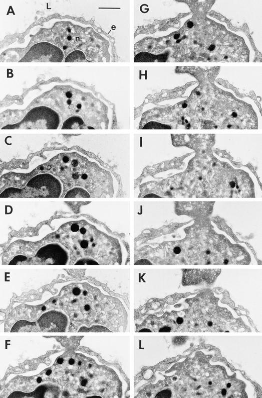
Transmigration of a neutrophil (n) across a thinned portion of venular endothelium (e) at a skin site injected 60 min earlier with 10−5 M FMLP. 12 of a series of 45 consecutive serial sections (sections 10–19 and 21–22) are illustrated and together encompass in its entirety the transendothelial pore (F–J) through which the neutrophil is migrating. Maximum pore diameter was 0.75 μm. L, lumen. Bar, 1 μm.