
Figure 7.
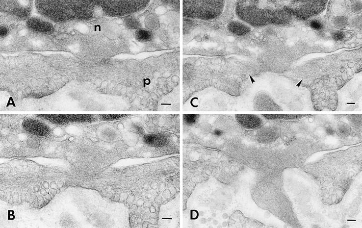
Neutrophil (n) migrating through a subendothelial pericyte (p) 60 min after intradermal injection of 10−5 M FMLP. Four (sections 12–14 and 16) of a series of 46 consecutive serial sections illustrate a transcytoplasmic pore in the pericyte through which the neutrophil has extended a blunt cytoplasmic projection. The projecting neutrophil pseudopod is filled with a feltwork of microfilaments. Note also the prominent, horizontally arrayed microfilaments in the immediately adjacent pericyte cytoplasm (C, arrowheads). Bar, 0.1 μm.