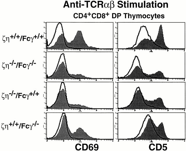
Figure 2.
CD69 and CD5 upregulation on DP thymocytes in response to TCR engagement. DP thymocytes were purified and stimulated for 12–16 h on plates coated with either PBS or anti–TCR-β. Cells were then stained with anti-CD69 or anti-CD5 and analyzed by FCM. Shaded areas depict cells stained with anti-CD69 or anti-CD5 after anti–TCR-β stimulation. Solid lines depict cells stained with anti-CD69 or anti-CD5 after incubation in media without antibody stimulation.

