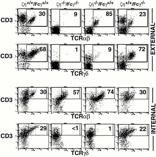
Figure 5.
i-IEL development in ζ/η−/−–Fcγ−/− mice i-IELs were prepared from mice as described (13) and three-color FCM was performed. For internal staining, cells were first stained with anti-CD4 and anti– CD8-β externally, then treated with intracellular staining buffer followed by staining with anti-CD3, anti–TCR-β, or anti–TCR-δ mAbs. Data depict two-color analysis of CD3 versus TCR-β or CD3 versus TCR-δ on software-gated CD4− CD8-β− cells. Numbers reflect the percentage of gated CD4−CD8-β− cells in that quadrant.

