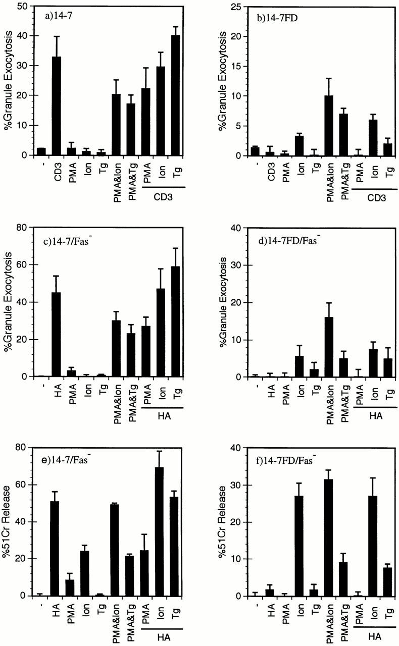
Figure 5.

A large increase in [Ca2+]i is required for perforin/granule exocytosis killing. 14-7 (a) and 14-7FD (b) were stimulated with plate-bound anti-CD3ε, PMA (25 ng/ml), ionomycin (Ion; 1,000 nM), thapsigargin (Tg; 200 nM), or a combination of PMA and ionomycin, PMA and thapsigargin, or anti-CD3ε plus PMA, ionomycin, or thapsigargin, respectively, in the absence of target cells, and percentage of granule exocytosis was determined. 14-7 (c, e) and 14-7FD (d, f ) were incubated with HA529–537 peptide (0.01 μM)–pulsed or mock-treated L1210Fas− target cells. At the start of the assay PMA, ionomycin, or thapsigargin were added at the aforementioned concentrations. After 4 h, supernatants were collected and percentage of granule exocytosis (c and d) and of specific killing (e and f ) was determined.
