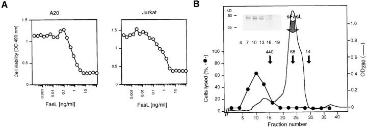
Figure 2.
(A) Characterization of FasL in supernatants of Neuro-2a cells transfected with muFasL. Crude supernatants were assayed for FasL activity on mouse A20 B cells and human Jurkat T cells. (B) Gel filtration analysis of FasL. Supernatants were concentrated and loaded onto a column (Superdex 200; Pharmacia). Fractions were analyzed for FasL by Western blotting in the presence of SDS (inset), for cell death–inducing activity (closed circles), and for protein (lines). Molecular size markers and the position of sFasL elution are indicated.

