
Figure 3.
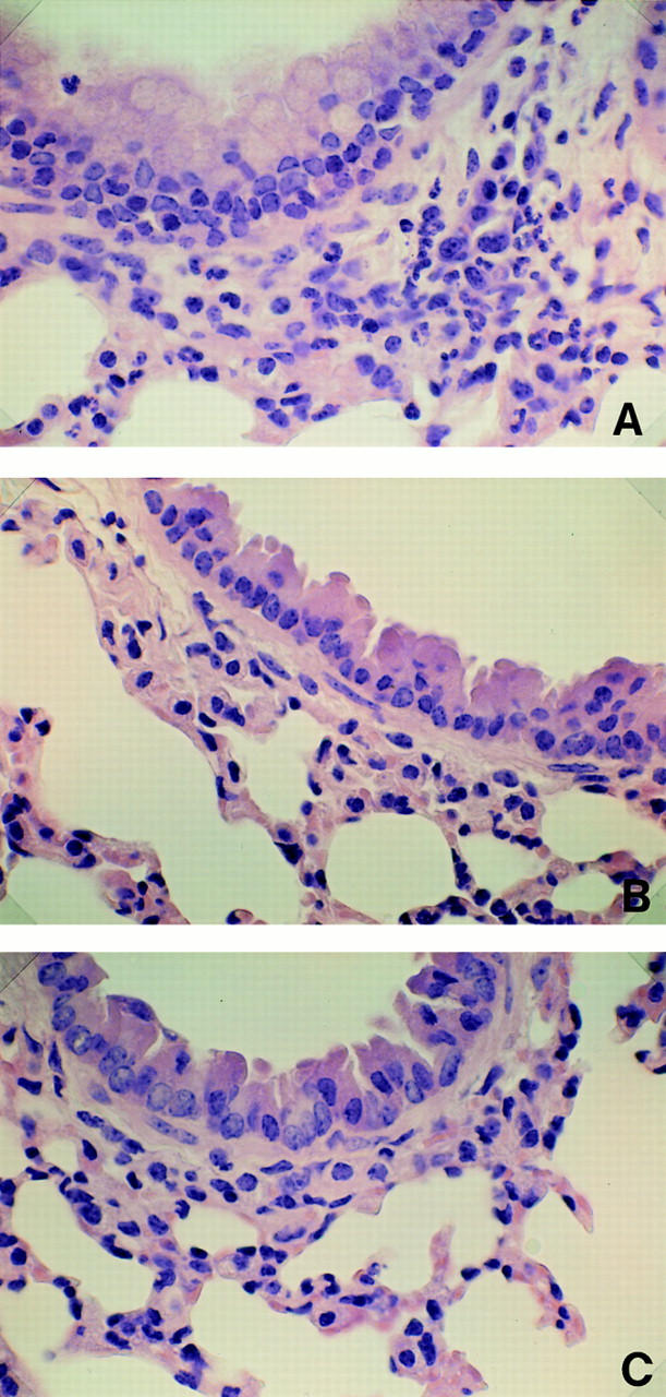
Microscopic examination of lung tissues from wild-type and STAT6−/− mice. Lung preparations from both wild-type and STAT6−/− mice treated with or without OVA were stained with hematoxylin-eosin. Photomicrographs of these preparations were evaluated infiltration of eosinophils and other lymphocytes in bronchial mucosa and perivascular sheaths in lung. Noticeable cell accumulation of eosinophils appeared in lungs from OVA-treated wild-type mice (A). In contrast, the inflammatory cells were not shown in lungs from OVA-treated STAT6−/− mice (B). The lungs taken from untreated wild-type mice represent normal lung histology (C). Original magnification: 200.