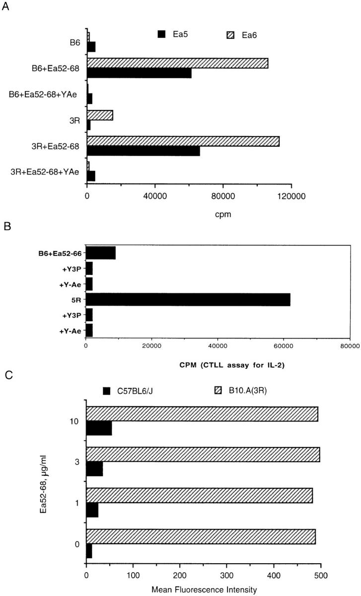
Figure 1.

Eα6 responds to Eα52–68 presented by I-Ab but not to endogenous ligand on B10.A (3R), whereas control hybrid 1H3.1 responds to both exogenous and endogenous ligand. (A) The proliferative response of the T cell clone Eα6 to APCs which did (3R) or did not (B6) express endogenous ligand, in the presence or absence of the peptide Eα52–68. The ability of the monoclonal antibody Y-Ae, which also recognizes the I-Ab–Eα peptide complex, to block the response of the clone is also shown. (B) The response of 1H3.1 was measured by culture of 105 hybrids with 2 × 10⁵ splenocytes along with the indicated antibodies and peptide. Culture supernatant was removed after 20 h and tested for the presence of IL-2 using the IL-2–dependent cell line CTLL. In A, the response to B6 splenocyte + the synthetic peptide Eα52–66 is shown. Responses to Eα52–68 were similar. In B, the response of 1H3.1 cells to the endogenous ligand on 3R cells is also shown. No synthetic peptide was added since 1H3.1 responds to the endogenous ligand. The antibodies were added to the culture wells to show that the response of 1H3.1 is restricted by I-Ab and is also blocked by Y-Ae. Y3P is an anti–I-Ab antibody. Y-Ae did not block the response of a control clone D10 (data not shown), which recognizes an I-Ab ligand different from that recognized by Y-Ae. Y-17, an anti–I-E antibody, does not block the response of either 1H3.1 or Eα6 (data not shown). (C) Flow cytometric staining with the antibody Y-Ae. Staining of B6 and 3R splenocytes in the presence of a titration of the peptide Eα52–68 is compared.
