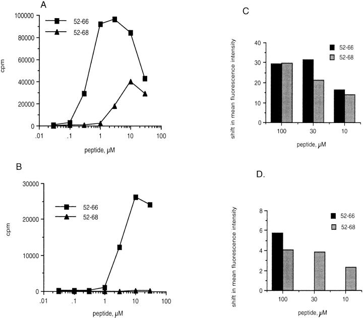
Figure 3.
Eα6 responds to Eα52–66 and Eα52–68 on live APCs, but does not respond to the longer peptide presented by fixed APCs. (A) Live APCs. The proliferative response of Eα6 to length variants of the Eα peptide presented on irradiated splenic APCs that could still take up antigen for processing (Live). (B) Fixed APCs + cross-linked anti-CD28. A similar proliferation assay, except the APCs were paraformaldehyde fixed splenocytes, which could no longer take up and process antigen. Anti-CD28 and goat anti–hamster IgG (Caltag Labs.) were added to the experiment in B to provide costimulatory signals, which could not be provided by the fixed APCs. C shows Y-Ae flow cytometric staining of live APCs after peptide loading, and D shows Y-Ae staining of APCs that were fixed before loading peptides. Y-Ae detects the loading of Eα peptides on I-Ab. The mean fluorescence intensity of unstained cells was subtracted from all samples to give a shift in mean fluorescence intensity. The mean fluorescence intensity of B6 APCs stained with Y-Ae in the absence of added Eα peptide was subtracted from the mean fluorescence intensity of samples stained after peptide loading in order to control for background staining by Y-Ae, which does not normally stain B6 APCs. The staining was performed in triplicate.

