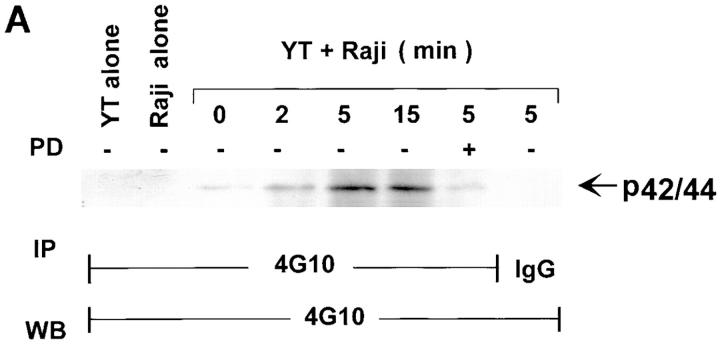
Figure 3.
Detection of tyrosine-phosphorylated p42/p44 MAPK protein in Raji-activated YT effector cells. (A) YT cells were cultured alone or with Raji cells at a 1:1 ratio for 0–15 min at 37°C. YT cells were also pretreated with 100 μM of PD098059 for 1 h at 37°C before incubation for 5 min at 37°C with Raji tumor cells. The cells were then lysed and immunoprecipitated (IP) with monoclonal antiphosphotyrosine, 4G10. Immunoprecipitation of YT cells, which had been preincubated with Raji tumor cells for 5 min at 37°C, with isotype-matched IgG was also performed as a control. Raji cells alone were included to check for background phosphorylation. The immunoprecipitates were then probed with 4G10 by Western blotting (WB). (B) YT cells were untreated or pretreated for 1 h at 37°C with 100 μM of PD098059 or an equivalent amount of DMSO used to dilute PD098059. The cells were then mixed with Raji tumor target cells for 0–5 min at 37°C and lysed. The lysates were immunoprecipitated with antiphosphotyrosine, 4G10, or control isotype-matched IgG and then probed with anti-panERK.


