Figure 4.
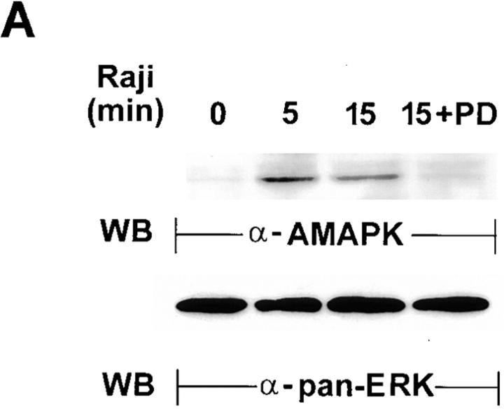
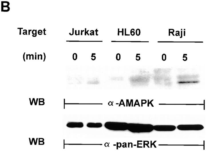
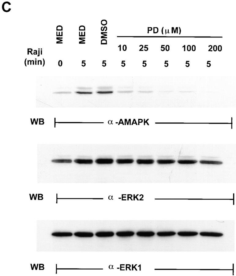
Detection of the activated form of MAPK in Raji-triggered YT effector cells. (A) YT cells untreated or treated with 100 μM of PD098059 for 1 h at 37°C were mixed with equal numbers of Raji tumor cells for 0–15 min at 37°C. Whole cell lysates were then prepared and analyzed by Western blotting (WB) with anti-AMAPK (α-AMAPK) that was generated against the phosphorylated TEY epitope of MAPK (top). The blots were then stripped and reprobed with anti-panERK to show equal loading of all the lanes (bottom). (B) YT cells were mixed with equal numbers of either Jurkat or HL60 tumor cells, both of which are NK-resistant, or with the NK-sensitive Raji tumor cells, for 0 or 5 min at 37°C. Whole cell lysates were prepared and analyzed by Western blotting with anti-AMAPK (top). The blots were then stripped and reprobed with anti-panERK (bottom). (C) YT cells were pretreated with medium (MED), DMSO, or 10–200 μM of PD098059 for 1 h at 37°C before addition of Raji tumor cells for 0 or 5 min at 37°C. Cell lysates were then prepared and probed with anti-AMAPK (top) and consecutively restripped and reprobed with anti-ERK2 (middle) and anti-ERK1 (bottom).


