
Figure 5.
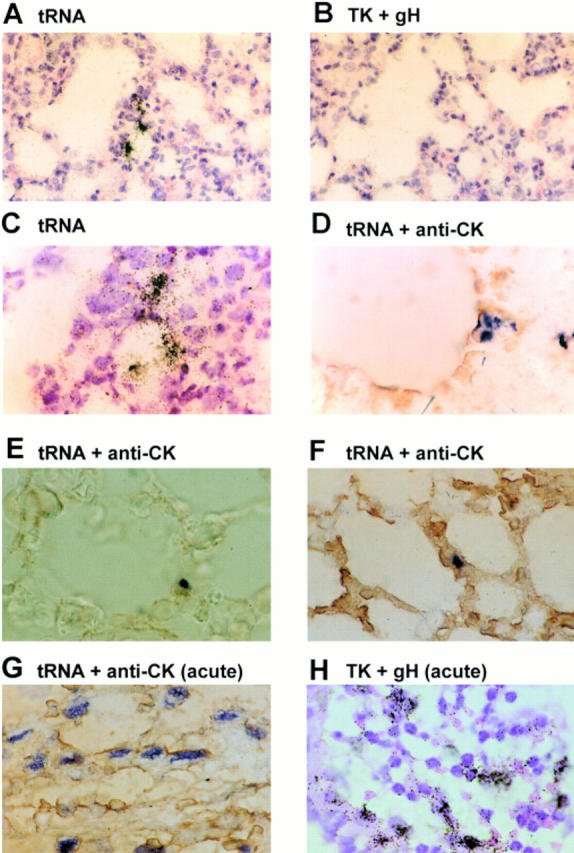
Determination of cell type harboring latent MHV-68 in the lungs by in situ hybridization. Sections of 5 μM thickness were cut from formalin-fixed paraffin-embedded lung tissues from mice during virus persistence at 54 d p.i. (A–F). Control sections were derived from mice at 5 d p.i. (G and H, acute). Sections shown in A–C and H were hybridized with 35S-labeled riboprobes and developed using photographic emulsion. The sections shown in D–G were hybridized with digoxigenin-labeled riboprobes and developed using a combination of alkaline phosphatase–labeled antidigoxigenin followed by NBT/BCIP (blue/black). Probes used were specific for either the latency-associated tRNAs (A and C–G, tRNA) or the productive cycle–associated thymidine kinase (TK) and gH genes (B and H, TK + gH). D–G were additionally reacted with anticytokeratin antibody and developed using DAB (brown, anti-CK). A and B are adjacent serial sections from one μMT mouse biopsy. All other panels are derived from different μMT mice. A–C and H were counterstained with hematoxylin and eosin. A and B, ×230; C–H, ×580.