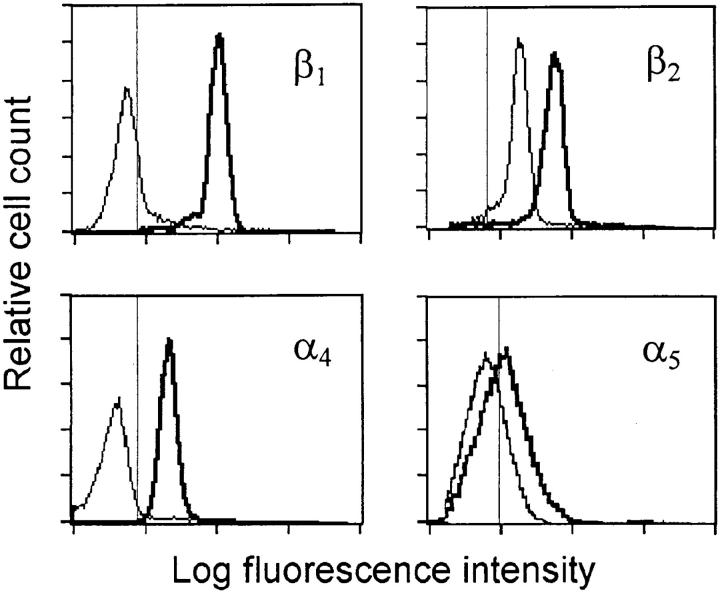
Figure 1.
Immunofluorescent staining of integrins on blood PMNs (thin line) and on extravasated PMNs collected from the peritoneal cavity (thick line). Thin vertical line indicates the 99th percentile of fluorescence events for cells stained with isotype matched control antibody. Histograms are representative tracings of three to five analyses for each antibody.

