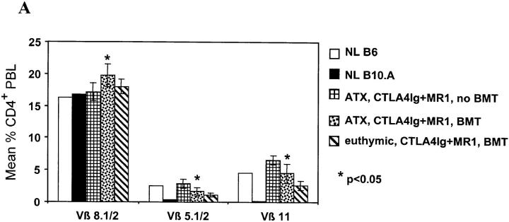
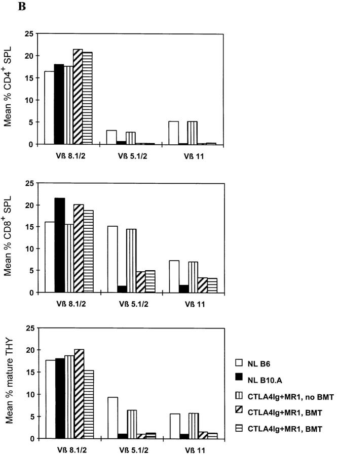
Figure 4.
Extrathymic clonal deletion after BMT and costimulatory blockade with CTLA4Ig and MR1. (A) ATX recipients (n = 6) showed specific, partial deletion of Vβ5.1/2+ and Vβ11+ CD4 cells in PBLs, 1 wk after BMT plus CTLA4Ig and MR1 and 3 Gy WBI. A similar degree of deletion was observed in euthymic controls (n = 5) prepared with the same conditioning. CD4 cells in ATX controls receiving CTLA4Ig plus MR1 and WBI without BMT (n = 4) did not show deletion of these Vβ. The percentage of Vβ+ cells was determined by FCM analysis of gated CD4+ PBLs. P values are shown for comparison between ATX BMT recipients receiving CTLA4Ig plus MR1 and ATX non-BMT controls. (B) In two euthymic chimeras killed 20 wk after BMT (under cover of 3 Gy WBI, plus treatment with CTLA4Ig plus MR1), Vβ5.1/2+ and Vβ11+CD4+ splenocytes (SPL) were deleted to the same extent as in naive B10.A controls (top). In contrast, the percentage of Vβ5.1/2+ and Vβ11+CD8+ splenocytes was reduced compared to naive B6 mice, but was substantially higher than in naive B10.A mice (middle). Mature Vβ5.1/2+ and Vβ11+ thymocytes (THY) showed deletion comparable to B10.A at the same time (bottom). A control mouse receiving WBI and CTLA4Ig plus MR1 (but no BMT) showed no deletion in either splenocytes or thymocytes. The percentage of Vβ+ cells was determined by FCM analysis in gated CD4+ (or CD8+) 34-2-12–negative splenocytes, and in gated KH-95high (i.e., Db high)-thymocytes (or Dd high in the case of BIO.A controls; see Materials and Methods). NL B6 denotes naive C57BL/6 control; NL B10.A denotes naive B10.A control.


