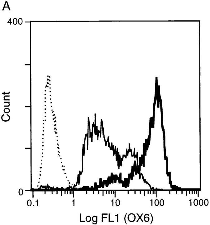
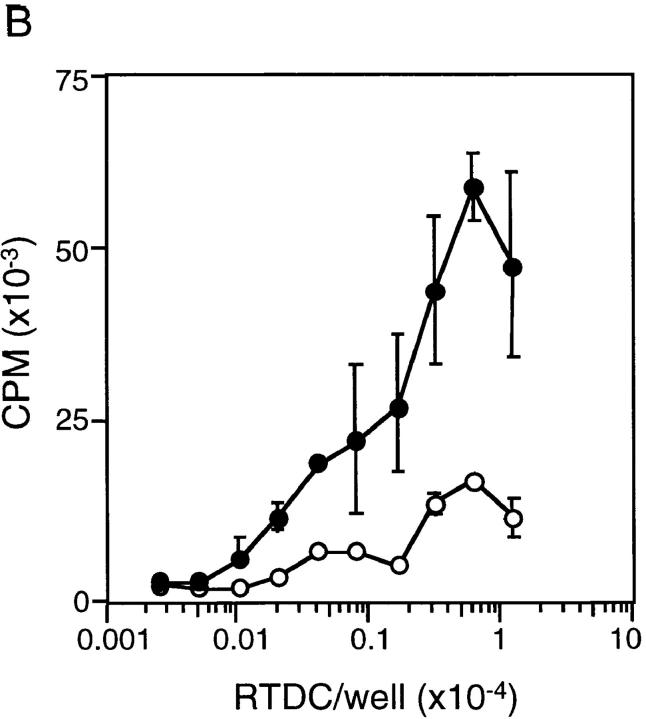
Figure 1.
Surface MHC class II expression and MLR-stimulating activity of fresh and GM-CSF–exposed RTDCs. (A) Purified RTDCs were labeled with OX6-FITC either as fresh cells (thin line) or after culture in GM-CSF (thick line), or with the isotype control IgG1-FITC (dotted line), and surface fluorescence analyzed by flow cytometry. (B) Serial dilutions of freshly purified (open circles) or GM-CSF–exposed (filled circles) RTDCs were added as stimulators to a primary allogeneic MLR in the presence of 105 purified WAG-strain T cells per well of 96-well microtiter plates. Cell proliferation (CPM) was assessed after a total of 96 h including an 18-h [3H]TdR pulse. Mean ± SEM of triplicate wells of one representative of three experiments is shown.


