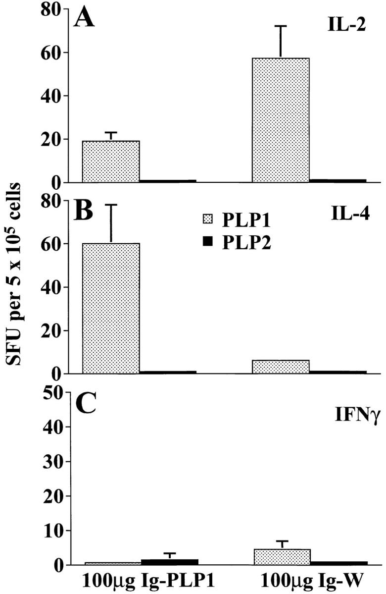
Figure 5.

Lymph node T cell deviation in mice recipient of Ig–PLP1 at birth. Newborn mice (eight per group) were injected intraperitoneally within 24 h of birth with 100 μg Ig–PLP1 or Ig–W in saline. When the mice reached 7 wk of age, they were immunized with 100 μg free PLP1 peptide in 200 μl CFA/PBS (vol/vol) subcutaneously in the foot pads and at the base of the limbs and tail. 10 d later the mice were killed, and the lymph node cells (0.4 × 10⁶ cells/well) were in vitro stimulated with free PLP1 or PLP2 (15 μg/ml) for 24 h. The production of IL-2 (A), IL-4 (B), and IFN-γ (C) was measured by ELISPOT as described in the Materials and Methods section using PharMingen anticytokine antibody pairs. The indicated values (spot forming units, SFU) represent the mean ± SD of eight individually tested mice.
