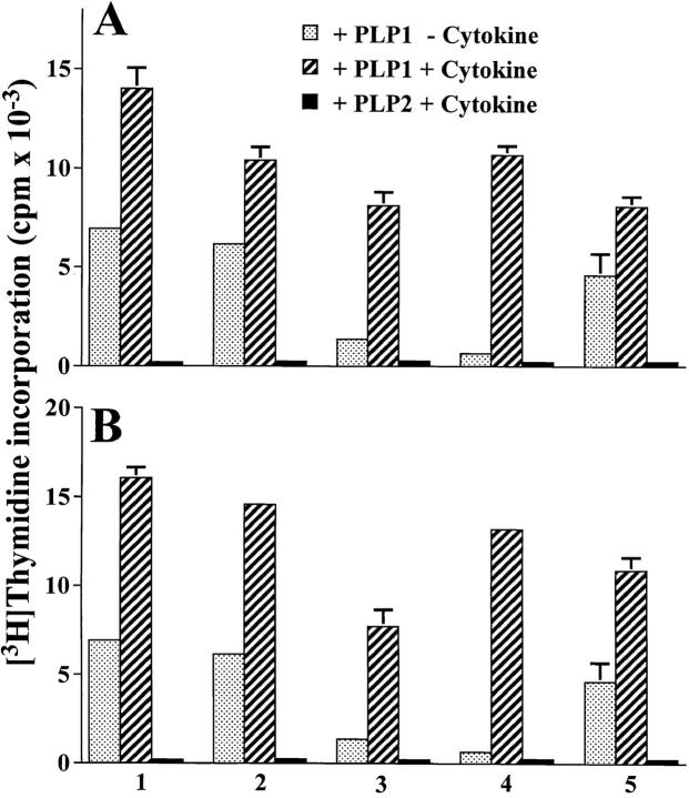
Figure 7.
Cytokine-mediated restoration of splenic T cell proliferation in mice injected with Ig–PLP1 at birth. A group of five newborn mice was injected intraperitoneally with 100 μg of Ig–PLP1 and immunized with 100 μg PLP1 peptide in CFA at 7 wk of age, as in Fig. 3. 10 d later, the splenic cells (106 cells/well) were in vitro stimulated with free PLP1 peptide (15 μg/ml) in the presence of 100 U/ml IFN-γ (A) or 10 U/ml IL-12 (B), and [3H]thymidine incorporation was measured as in Fig. 3. Cells from each mouse were stimulated with PLP1 peptide without addition of exogenous cytokines (dotted bars), with PLP1 peptide in the presence of cytokine (hatched bars), or with PLP2 peptide in the presence of cytokine (black bars). The indicated cpms for each mouse represent the mean ± SD of triplicate wells.

