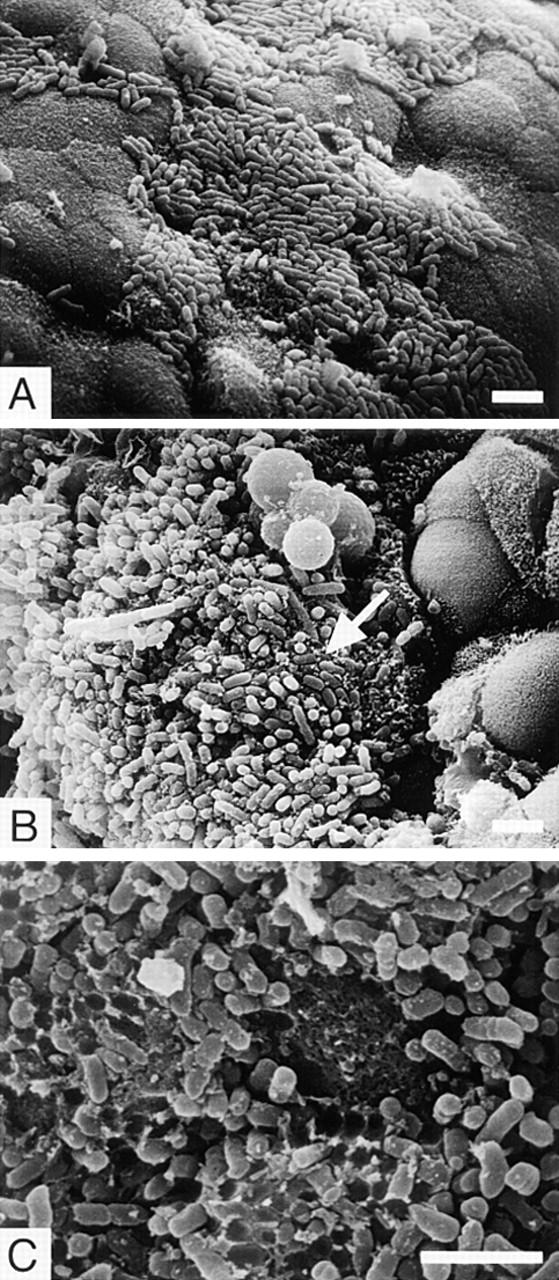
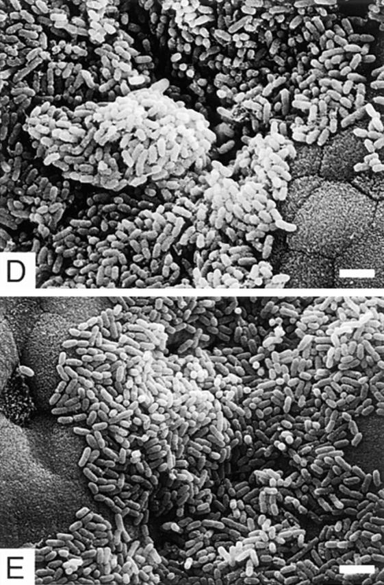
Figure 5.


Scanning electron micrographs of the ileal villi infected with REPEC O103 (A–C), AAF004 (D), and AAF005 (E). REPEC O103, AAF004 (EspA−), and AAF005 (EspB−) adhered to the villi, and the adherence pattern is similar between wild-type and mutant strains. However, embedded bacteria as indicated by an arrow (B) and cup-like structures (C) were observed only in rabbits infected with REPEC O103. Ileal sections were taken 72 h (for REPEC O103 and AAF005) and 96 h (for AAF004) after infection. Bar, 4 μm.
