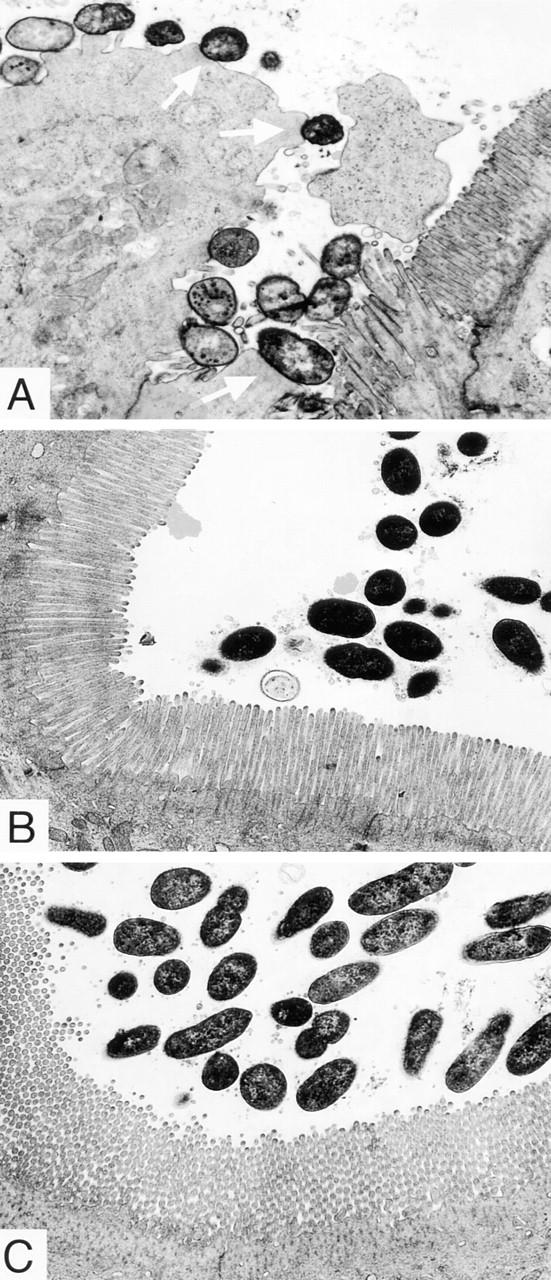
Figure 6.

Transmission micrographs of the ileal villi infected with REPEC O103 (A), AAF004 (B), and AAF005 (C). The REPEC O103 are intimately associated with the ileal villi and form pedestal-like structures, which are indicated by arrows. Microbodies and elongated and swollen microvilli were also observed. In contrast, AAF004 (EspA−) and AAF005 (EspB−) do not cause A/E lesions, and intracellular damage was not seen. All ileal sections were taken 96 h after infection. Original magnification, ×6,300.
