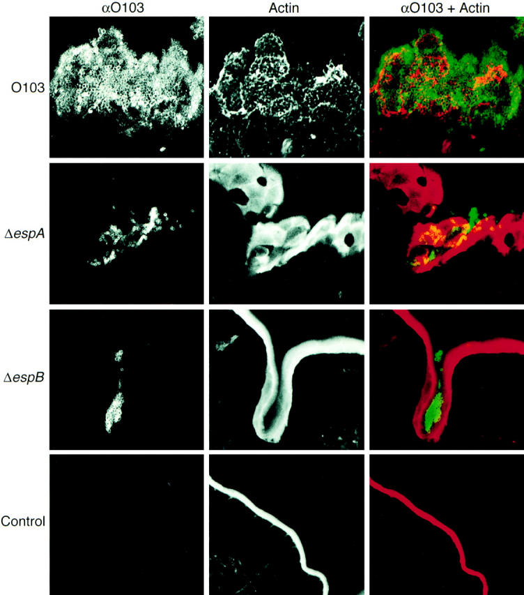
Figure 7.

Confocal laser scanning micrographs of Peyer's patches infected with REPEC O103, AAF004, and AAF005. Peyer's patches were taken 72 h after infection and cryosections (20-μm-thick) were stained with phalloidin-TxR (red for overlay) and anti-O103 antibody (green for overlay). REPEC O103 adhered and colonized to the Peyer's patches, cytoskeletal actin beneath the attached bacteria was rearranged, and cup-like structures were also observed. In contrast, a small number of EspA and EspB mutant strains were observed in villi crypts, but no actin rearrangements occurred. Strains: REPEC O103, O103; AAF004 (EspA−), ΔespA; AAF005 (EspB−), ΔespB; PBS inoculation, Control.
