
Figure 7.
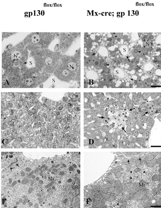
Hepatic abnormalities in the conditional mutants. Light (A–D) and electron (E and F) microscopic analysis of control (A, C, and E) and gp130-mutant (B, D, and F) liver. N, Nucleus; S, liver sinus. After 6 wk, gp130-mutant liver (B) has a high amount of lipid droplets (arrowheads), widening of the Disse and intercellular space (arrows), a reduced content of binucleated hepatocytes, and an increase in Kupffer cells compared with controls (A). In 12-mo-old gp130-mutant liver, a part of the liver parenchyma is replaced by fibrotic material (D, arrows), a phenomenon not observed in the gp130flox/flox control animals (C). The ultrastructure of gp130-mutant liver (F) gives evidence for a rarification of smooth and rough endoplasmic reticulum (ER), a dense clustering of mitochondrial (Mi) and monoparticulate (β) glycogen granules (triangle), a decrease of microvilli (*), and an enlargement of Disse and intercellular spaces (arrows) compared with the gp130flox/flox control (E). For A and B, bar = 5 μm; for C and D, bar = 20 μm; for E and F, bar = 0.5 μm.