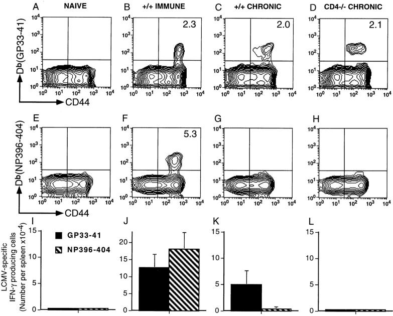
Figure 2.
T cell deletion and functional unresponsiveness are distinct mechanisms for silencing antiviral immune responses. Naive +/+ mice, +/+ immune mice (161 d after LCMV Armstrong infection; 2 × 10⁵ PFU, i.p.), and +/+ or CD4−/− mice infected with LCMV-t1b (2 × 10⁶ PFU, i.v.) 60 d previously were checked for the physical presence and functional responsiveness of LCMV-specific CD8 T cells. (A–H) LCMV-specific CD8 T cells were visualized using MHC class I tetramers complexed to viral peptides. Cells were costained with anti-CD8–PE, anti-CD44–FITC, and either Db(GP33–41) or Db(NP396–404) tetramers conjugated to allophycocyanin. The histograms show gated CD8 lymphocytes, and the percentage of CD8 cells costaining with either Db(GP33–41) or Db(NP396–404) are indicated in the corresponding upper right quadrant. Where not shown values are <0.2%. (I–L) Numbers of LCMV-specific IFN-γ producing cells were enumerated using single-cell cytokine ELISPOT assays. The number of splenocytes producing IFN-γ after stimulation with either GP33–41 (black bars) or NP396–404 peptides (hatched bars) are shown (± SD).

