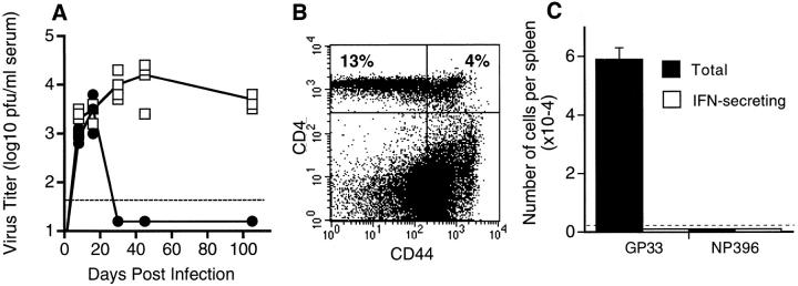
Figure 4.
Chronic LCMV infection and CD8 T cell unresponsiveness ensue after transient loss of CD4 T cells. +/+ mice were transiently depleted of CD4 T cells by injection with the anti-CD4 antibody, GK1.5, 1 d before and 3 d after infection with LCMV-t1b. (A) Serum virus titers were determined in +/+ (•) and GK1.5 treated (□) +/+ mice at various days after infection. (B) Reconstitution of CD4 T cells in GK1.5-treated mice was determined at 60 d after infection with LCMV-t1b. Splenocytes were costained for CD4 and the activation marker CD44. The percentage of CD4+ cells are indicated in the upper quadrants. By this time point, both +/+ and GK1.5-treated mice contained comparable numbers of CD4 T cells. (C) Splenocytes from GK1.5-treated mice were prepared at 60 d after infection with LCMV-t1b and both the total number (measured by tetramer staining) and the number of IFN-γ–secreting (measured by ELISPOT) GP33- or NP396-specific CD8 T cells were determined. Greater than 98.5% of GP33-specific T cells were unresponsive and NP396-specific T cells were not detectable. In A and C the limit of detection is indicated by the dashed line. At least three mice were analyzed at each time point.

