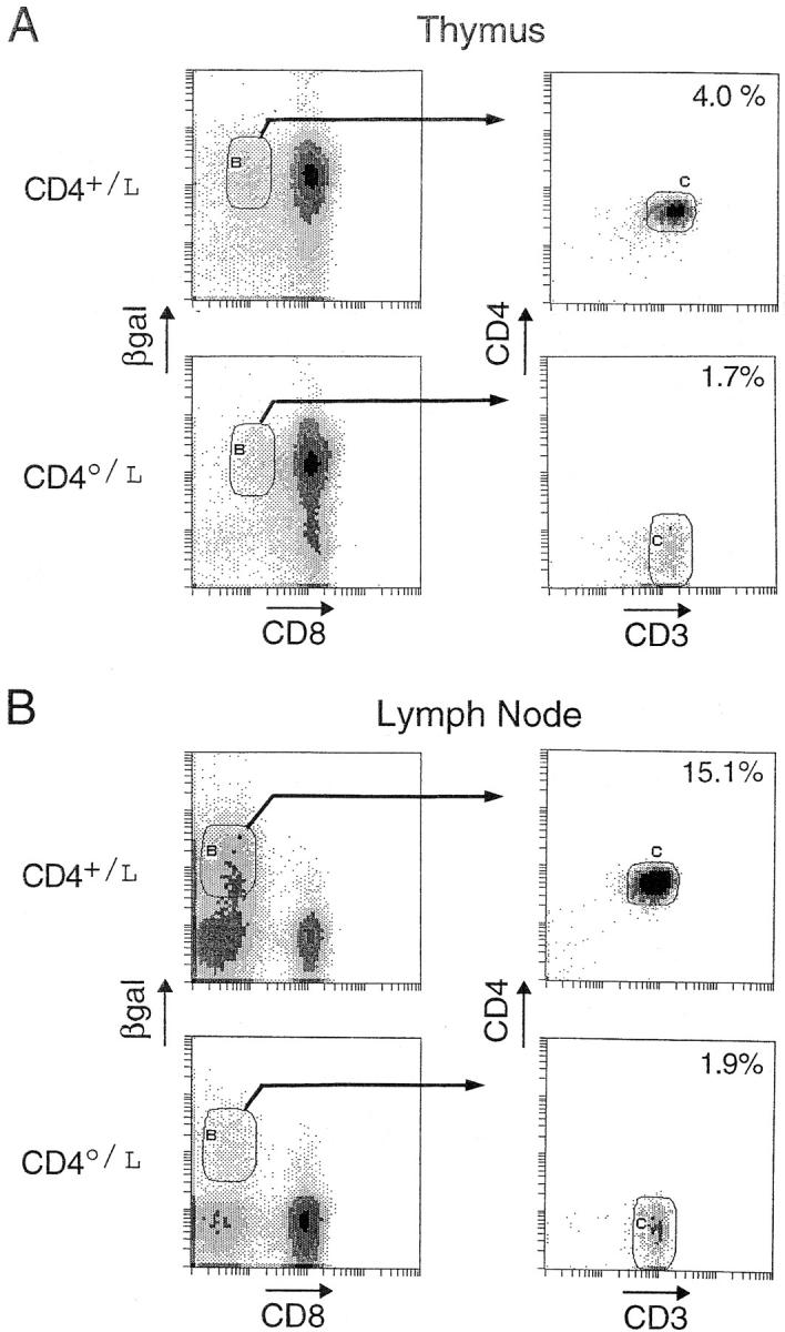
Figure 9.

βgal+ DN cells in CD4-deficient mice. Thymocytes and lymph node cells from homozygous (CD40/L) and heterozygous (CD4+/L) mice were compared using four-color flow cytometry. Cells were first plotted for their expression of βgal and CD8. CD8+βgal+ cells were gated and further evaluated for their expression of CD4 and CD3. Percentages are of total thymocytes.
