Figure 1.
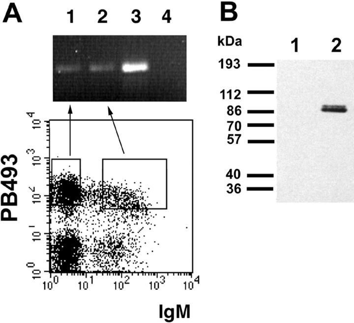
Expression of Egr-1 during different stages of B cell maturation. (A) Egr-1 RNA expression. Bone marrow cells of a 5-wk-old BALB/c mouse were stained for B220, PB493, and IgMa expression. Pro/pre-B cells (B220low, PB493+, and IgM−) and immature B cells (B220low, PB493+, and IgM+) were sorted and RNA was extracted. Analysis of Egr-1 transcripts by reverse transcription PCR was performed as described by Miyazaki (35). Lane 1 shows expression of Egr-1 in pro/ pre-B cells and lane 2 in immature B cells. Anti-IgM–stimulated splenocytes (lane 3) serve as a positive control. In lane 4, cDNA was omitted from the PCR. (B) Expression of Egr-1 protein. BALB/c fetal liver B cells (day 16) were cultivated in the presence of IL-7 on ST-2 stroma cells. Cellular lysates of 106 cells were examined for Egr-1 expression by immunoblotting using the Egr-1–specific antibody C19 and developed with horseradish peroxidase–coupled goat anti–rabbit IgG. In cultivated pre-B cells, Egr-1 protein expression is easily detected (lane 2). As a negative control an equal amount of ST-2 feeder cells was loaded in lane 1.

