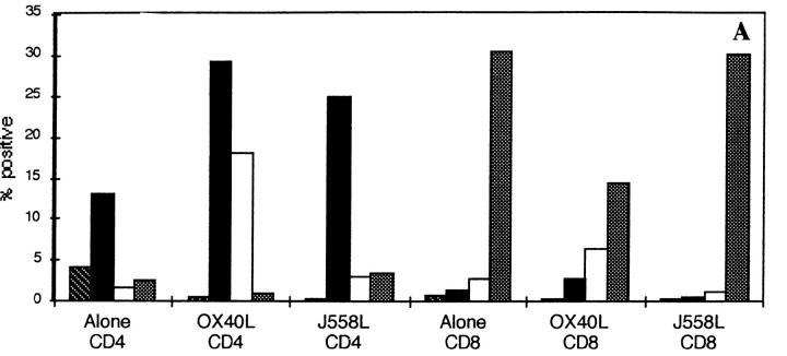
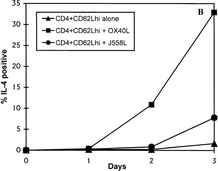
Figure 3.
Induction of IL-4 expression by OX40L. CD62Lhigh CD8 and CD4 T cells were activated through CD3 and CD28 for 3 d in the presence of a fixed OX40L transfectant or the control parental cell line. Cells were restimulated through CD3 for 4 h in the presence of GolgiStop™ and stained for intracellular expression. (A) Staining with a control mAb (striped bars), anti-CD40L (black bars), anti–IL-4 (white bars), and anti–IFN-γ (stippled bars), on purified CD4 or CD8 CD62Lhigh T cells. The above data are representative of at least 10 different experiments. (B) Timing of induction of IL-4 expression in naive T cells by OX40L. Cells were stimulated as above. Data are representative of two experiments. hi, High.


