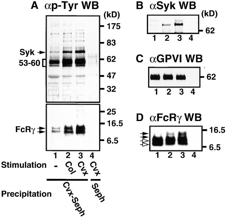
Figure 2.
Tyrosine phosphorylation of proteins coprecipitated with the GPVI–FcRγ complex in collagen- and convulxin-stimulated platelets. Gel-filtered platelets were unstimulated (lane 1) or stimulated for 1 min at 37°C with 50 μg/ml of collagen (lane 2) or 50 ng/ml of convulxin (lanes 3 and 4) and lysed in Triton X-100 lysis buffer. Precipitated proteins with convulxin-coupled Sepharose 4B (lane 1–3) or Sepharose 4B (lane 4) were resolved by 10% (A, top, B, and C) or 12.5% SDS-PAGE (A, bottom and D), transferred to nitrocellulose membranes, and immunoblotted with Abs against phosphotyrosine (A), Syk (B), GPVI (C), and FcRγ (D). Molecular mass markers are indicated in kD on the right of panels. In A (bottom) and D open and closed arrows mark unphosphorylated and phosphorylated FcRγ, respectively.

