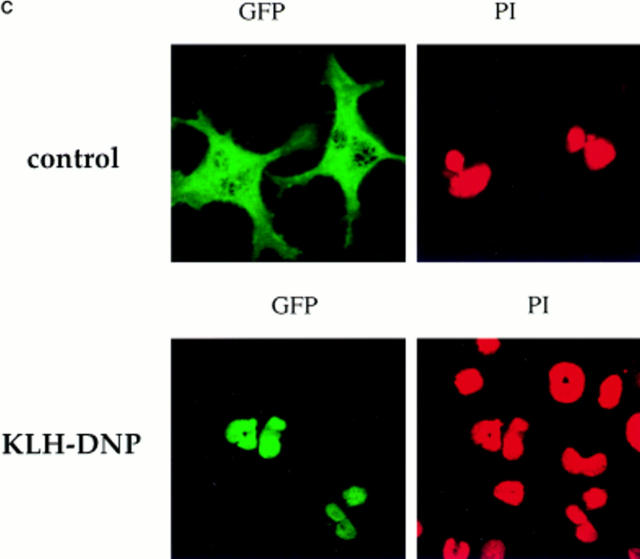
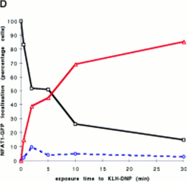
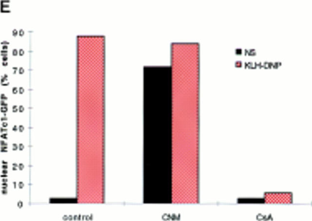
Figure 1.
(A) NFATC1-GFP marker construct. Schematically shown here, the full-length NFATC1 molecule was fused to the GFP reporter by cloning into the pEGFP-1 vector (Clontech). TAD, Transactivation domain. HR, Homology region. SRR, Serine-rich region. SP, Ser/Pro box. (B) NFATC1-GFP is cytosolic in resting RBL2H3 mast cells. RBL2H3 cells were transfected with 8 μg NFATC1-GFP and recovered for 6 h on glass coverslips in complete medium at 37°C. Cells were costained with PI, fixed, and mounted as described. (C and D) FcεR1 cross-linking induces NFATC1-GFP nuclear import in RBL2H3 mast cells. RBL2H3 cells were transfected with 8 μg NFATC1-GFP and recovered for 6 h on glass coverslips in complete medium at 37°C. Cells were IgE primed and stimulated with 500 ng/ml KLH-DNP for 30 min (C) or for the indicated times (D). Cells were either costained with PI, fixed, and mounted as described (C), or cells were scored for predominant localization of GFP (D). (E) FcεR1 induction of NFATC1-GFP nuclear import is CN regulated and CsA sensitive. RBL2H3 cells were transfected with 8 μg NFATC1-GFP reporter alone or in combination with 20 μg activated CN plasmid (CNM) and recovered for 6 h on glass coverslips in complete medium at 37°C. Cells were primed and stimulated with 500 ng/ml KLH-DNP for 30 min in the absence or presence of 50 nM CsA. NS, Not stimulated. Cells were fixed, mounted, and scored for predominant localization of GFP as described.


