
Figure 1.
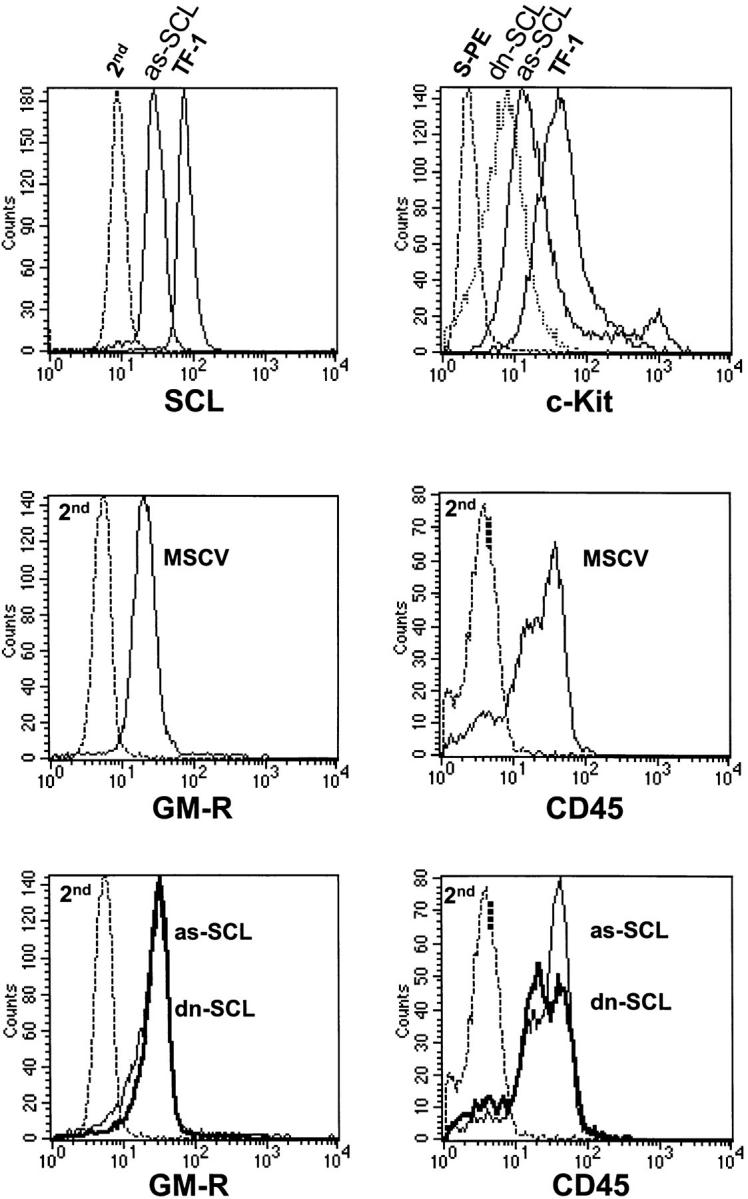
Flow cytometry analysis of TF-1 transfectants expressing as-SCL or dn-SCL. (top left) Decreased SCL protein in TF-1 cells expressing as-SCL. TF-1 cells were infected with a retrovirus harboring the human SCL cDNA cloned in the antisense orientation and maintained as a polyclonal population in GM-CSF containing medium in the presence of G418 (1 mg/ml). 1 wk after infection, as-SCL transfectants were fixed and permeabilized with Triton X-100 for nuclear staining with the monoclonal anti-TAL1 at a final dilution of 1:10, followed by labeling with a goat anti–mouse antibody coupled to FITC (Sigma). Cells were washed extensively for 10 min after each labeling step and analyzed by flow cytometry. Parental TF-1 cells were stained in parallel as positive controls. Both cell types were also stained with the second antibody alone (2nd) as negative controls. In contrast to the shift in SCL nuclear staining between as-SCL transfectants and parental TF-1 cells, those of MSCV (vector alone) transfectants and TF-1 cells were comparable (data not shown). (top right) Decreased c-Kit expression in TF-1 cells expressing as-SCL or dn-SCL. The same cells as in the top left panel were analyzed for surface labeling with a biotinylated monoclonal antibody against human c-Kit and streptavidin-coupled PE (S-PE). Cells labeled with S-PE alone serve as negative controls. (middle and bottom) Surface expression of CD45 and CD116 (GM-CSF receptor [GM-R] α chain) is unaltered in TF-1 transfectants. Cells were labeled with a mouse anti–human CD45 and anti–human CD116 (GM-R), followed by FITC-labeled goat anti–mouse IgG. Labeling with the second antibody alone (2nd) is shown.