
Figure 9.
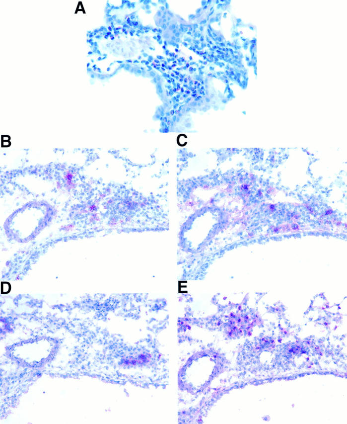
Characterization of inflammatory cells in lung sections from an IL-9–expressing mouse. Formalin-fixed tissue was stained with new vital red to demonstrate that eosinophils were present in high numbers in lung tissue from IL-9–expressing mice (A). Immunostaining of frozen lung sections (B–E) revealed that cell infiltrates also contained CD4+ (B), CD8+ (C), B220+ (D), and Mac-1+ (E) mononuclear cells. Original magnifications: A, ×600; B–E, ×500.