
Figure 1.
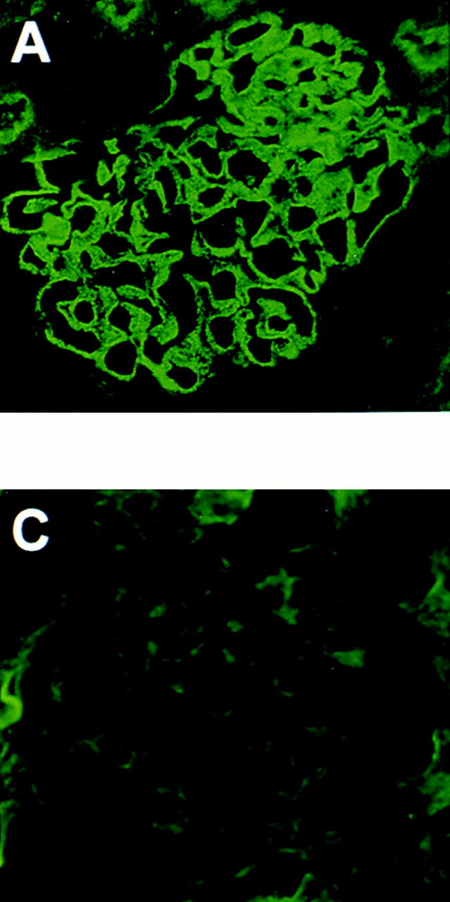
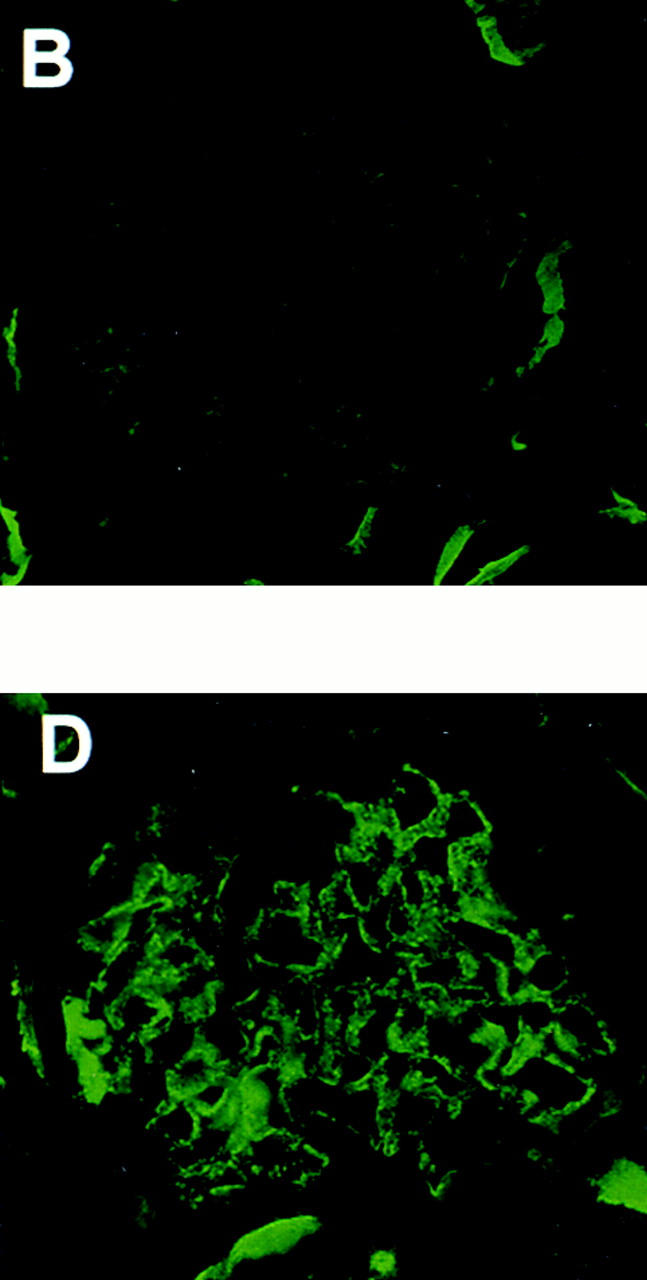
Representative IF micrographs in animals immunized with Fx1A lacking Crry. 10 wk after immunization, renal biopsies were performed and tissue was stained for IgG (A) and C3 (B). Animals were then injected with preimmune IgG (C) or anti-Crry IgG (D). 6 d later, animals were killed and renal tissue was stained for C3 (C and D). The IF micrographs shown in A, B, and D are from the same animal, and show typical granular staining for IgG in HN (A) and the appearance of a similar staining pattern for C3 only after injection of anti-Crry IgG (D).