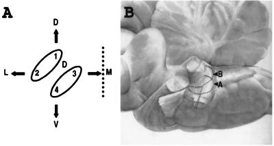
Figure 5.
(A) Schematic cross-section of the lateral line somatotopy in the hindbrain. The dotted line represents the midline. 1, 2, 3, 4, and D refer to the neuromasts labeled on Fig. 1B. The upper ellipse (1 and 2) represents the cross-section of the ribbon-like projection of the posterior lateral line; the lower ellipse (3 and 4) that of the anterior lateral line, D stands for the projection of the dorsal neuromast. Arrows: D, dorsal; V, ventral; M, medial; L: lateral. (B) Somatotopic projection of the spiral ganglion neurons in the cochlear complex of the cat. Side view, anterior to the left, dorsal to the top. The projection of hair cells located at the apex (A) of the Corti organ is ventrolateral to that of cells located more basalward (B). After ref. 1. Magnification: ×0.5.

