
Figure 4.
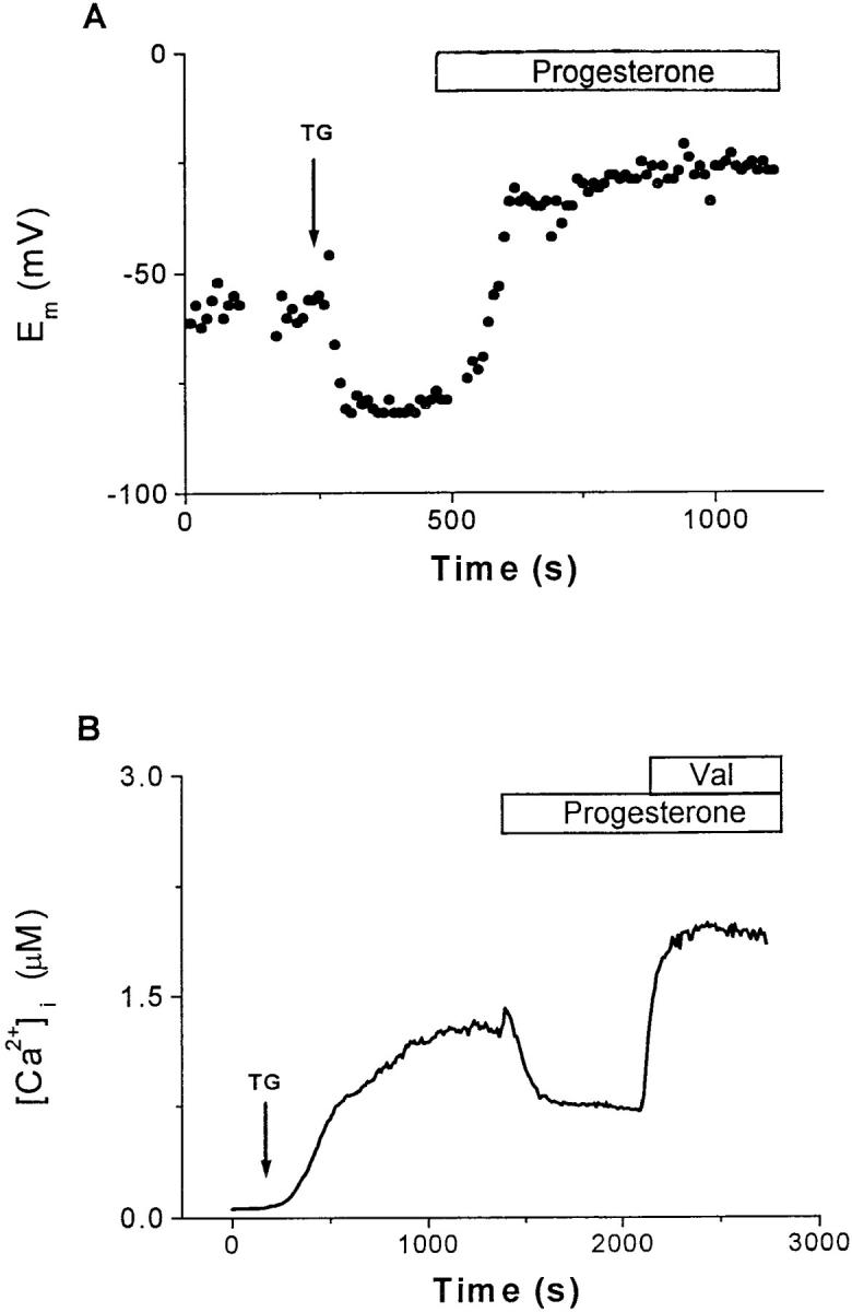
Progesterone reduces the driving force for Ca2+ influx. (A) Current-clamp recordings using the perforated-patch technique were performed in B3Z cells to determine the effects of progesterone on E m. This panel presents a representative single-cell recording. The addition of 1 μM TG (arrow) hyperpolarized the cell from the resting potential (−60 mV) to near the K+ equilibrium potential (∼−80 mV), enhancing the driving force for Ca2+ influx. Subsequent application of 50 μM progesterone depolarized the cell to −26 mV, resulting in a reduction of the driving force for Ca2+ influx. For perforated-patch recordings, the tips of the pipettes were filled with the following solution (in mM): 120 K2SO4, 16 KCl, 5 MgSO4, 10 Hepes (pH 7.2). A stock solution of nystatin in DMSO (25 μg/ml) was prepared daily and subsequently diluted in the pipette solution to a final concentration of 100 μg/ml. After sonication, this solution was used for backfilling the pipettes as described previously (reference 31). (B) Measurements of average [Ca2+]i from B3Z cells (n = 37). After the addition of 1 μM TG (arrow), [Ca2+]i rose from a resting level of 70 ± 30 nM to a plateau of 1.3 ± 0.4 μM. Progesterone (30 μM) reduced the [Ca2+]i to approximately half of plateau concentration, an effect that was completely reversed by the addition of 2 μM valinomycin. Application bars, The additions of progesterone and valinomycin to the bath.