
Figure 2.
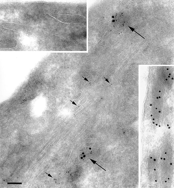
Immunoelectron microscopic colocalization of IL-8 and vWf in Weibel-Palade bodies. HUVEC were stimulated with 1,000 U/ml IL-1β overnight and processed for electron microscopy as indicated in Materials and Methods. Large granules (18-nm gold) represent vWf; small granules (6-nm gold) correspond to IL-8. Short arrows, IL-8 in the Golgi apparatus; long arrows, Weibel-Palade bodies that contain both IL-8 and vWf. Top left inset, The absence of granules from Weibel-Palade bodies stained with the irrelevant antibodies rabbit antioccludin and mouse anti–E-selectin. Bottom right inset, Weibel-Palade bodies in unstimulated HUVEC contained only vWf, but no IL-8. Scale bar, 100 nm.