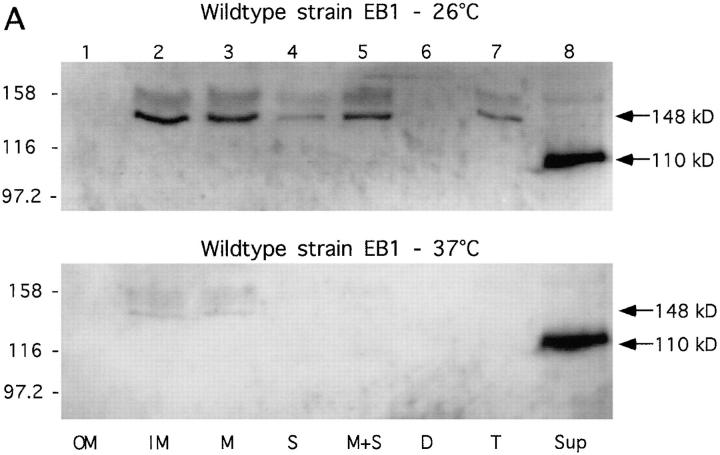
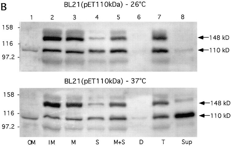
Figure 7.
Subcellular localization of Hbp. Cells of the wild-type strain EB1 (A) and IPTG-induced cells of strain BL21(pET-Hbp) (B) were grown in LB medium at 26°C and 37°C. Outer membrane (OM), inner membrane (IM) and crude membrane (M) fractions were derived from 1.3 OD660 units. Soluble (S), membrane and soluble (M+S), cellular debris (D), and total lysate (T) fractions were derived from 0.33 OD660 units. Culture supernatant (Sup) fractions were derived from 3.0 OD660 units. The fractions were analyzed by immunoblotting with the anti-Hbp antiserum. The secretory intermediates of Hbp and molecular mass markers (kD) are indicated at the right and left side of the panels, respectively.


