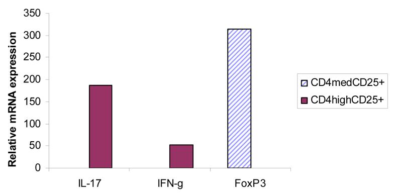
Figure 5. Inflammatory cytokine and FoxP3 gene in sorted cell subsets.
MOG peptide primed and in vitro stimulated LN cells were stained with CD4 and CD25 antibodies as described in methods. CD4high Teff cells and CD4normalCD25+ Treg cells were sorted using gating similar to that shown in Figure 3. Gene expression from sorted cells (8 × 10⁴ cells) was analyzed by real time PCR.

