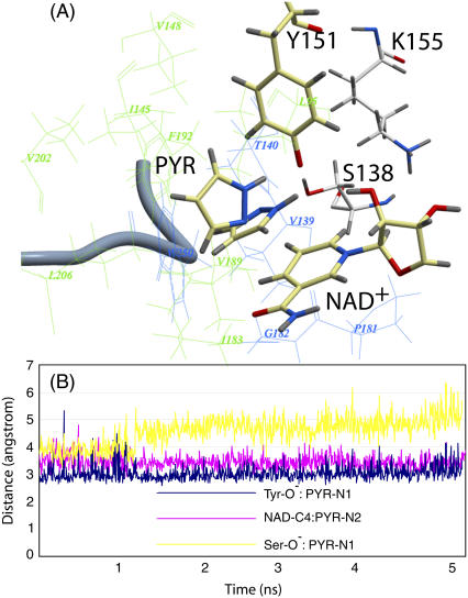
FIGURE 5.
Ternary DlADH·NAD+·PYR complex with the  ionization scenario of the catalytic triad after 5 ns of MD. (A) Amino acids lining the R1 (wire model in green) and R2 (wire model in blue) subpockets of the active site. The nicotinamide and ribose ring of NAD+, the catalytic triad (Ser-138, Tyr-151, and Lys-155), and PYR are shown as stick models with coloring according to the atom type (carbon-yellow, nitrogen-blue, oxygen-red, and hydrogen-gray). The ribbon represents the C-terminal loop of the other subunit acting as a lid of the active site cavity. PYR is shown both before and after the 5 ns of MD. The ring was parallel to the nicontinamide ring of the NAD+ ring at the start of the MD but tilted to perpendicular during the MD. (B) Fluctuation of atomic distances between PYR and atoms of the active site during the MD.

